Wednesday, March 16, 2011

Corneal Cross-linking Illumination System

Crosslinking with riboflavin and UV-A light has proven to be a first-line treatment for people with corneal ectasia disorders such as keratoconus, and pellucid marginal degeneration and corneal weakness (ectasia) after LASIK.

Photo-polymerisation with the use of ultra-violet light is the most promising technique to achieve cross-linking in connective tissue. Photo-polymerisation is activated by means of a non-toxic and soluble photomediator together with a wavelength of 365 nm UV-A light source.

A magnified section of the cornea showing all layers of the cornea in cross-section.
Fig 1, shows corneal crosslinking strengthens bonds in the stroma of the cornea, which is the layer from which tissue is removed during LASIK surgery


Fig 2, shows UV-X in use for eye treatment.


Homogeneous UV-A irradiation of the corneal is an important factor for a safe treatment procedure.Thus a homogeneous illumination of the cornea with a patented beam and optics is essential - the IROC UV-X. UV-X was developed by an experience team of researchers in collaboration with the inventors of the procedure, Prof Theo Seiler and Prof Eberhard Spoerl. It is designed with a special focus on both safety and effectivity of the procedure, embodying the cutting-edge knowledge in the field of corneal cross-linking.


Fig 3, shows Scanning Electron microscopy image of the homogenizing micro-structure used in the UV-X.


Pictures of the UV-X treatment versus the LED techniques are shown below:

Fig 4, shows UV-A versus direct LED irradiation

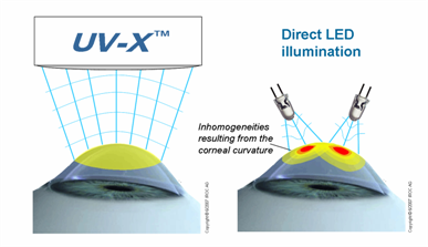
Fig 5, shows compensation of the corneal curvature using UV-X.

Fig 6, shows comparison of the treatment distance with reference to intensity-damage threshold.


Fig 7, shows an optical design according to Koehler's reduces the radiant exposure to retina.

Once patients have received crosslinking, in most cases their corneal shape will either remain stable or actually improve. This can provide patients with improved visual acuity and improved ability to wear contact lenses.

 Some surgeons have also reported reasonable results in improving the stability of the cornea in people who have undergone radial keratotomy, an incisional refractive procedure from the 1980s and 1990s. Crosslinking appears to work best for those who are experiencing daily fluctuations in their vision.

People who are considering vision correction procedures such as LASIK also might eventually be pre-treated with corneal crosslinking to strengthen the eye's surface before undergoing an excimer laser ablation that reshapes the cornea.


Source:  http://www.allaboutvision.com/conditions/corneal-crosslinking.htm


Wednesday, March 2, 2011

Fibroscan an alternative to liver biopsy

Fibroscan technique is a concept developed and produced by Echosens.

The FibroScan technique is used to quantify hepatic fibrosis in a totally non-invasive and painless manner, with no contra-indications for the patient. It is basically an ultrasound scanning system assessing liver scarring, termed fibroisis. Here the wave generated by the ultrasound transducer is propagated down to amd through the liver in which the velocity of the wave is measured. The speed of the wave reflects the degree of fibrosis and correlates with the stiffness of the liver. A higher degree of fibrosis correlates to higher stiffness of the liver. Fibroscan uses Kilopascals as the unit of measurement. The measurement is a median score of at least 10 readings. In viral hepatitis, a score of less than 7 means no or insignificant liver fibrosis. A score  of more than 12.5 KPa is severe fibrosis or cirrhosis and the results between this suggest moderate fibrosis.

Does FibroScan Accurately Assess Liver Fibrosis?

 Vibration-controlled transient elastography (VCTE) with FibroScan can provide an accurate assessment of liver fibrosis in patients with chronic viral hepatitis, but operator-related and patient-related factors affect measurements.
(Report 2 articles published in the April issue of Clinical Gastroenterology and Hepatology.) The lowest levels of variation occur in patients with no or early-stage fibrosis, or when the procedure is performed by a single experienced operator, the studies show.

It is important to determine the level of fibrosis in patients with chronic liver diseases, including nonalcoholic steatohepatitis (NASH), to evaluate disease progression. Until recently, liver disease progression could only be monitored by collection of biopsies and histologic analyses. However, liver biopsies provide information about only a small part of the liver, and are uncomfortable and even dangerous for patients.

VCTE with FibroScan (Echosens; Paris, France) transmits a vibration at low frequency to induce an elastic shear wave; the velocity of the wave propagation can be measured and directly relates to the tissue stiffness. VCTE acquires information from about 100-fold more sample tissue than a liver biopsy. Furthermore, the procedure is rapid, noninvasive, and can be performed at the bedside, providing the physician with immediate information for patients. Findings from VCTE are generally considered to be reproducible.

Information on this page is provided for interest only on a "best efforts" basis and does not 
constitute personal advice. Always discuss medical conditions and related matters with your doctor.

Reference: http://journalsblog.gastro.org/does-fibroscan-accurately-assess-liver-fibrosis/